Diagnostics
OCT
Imaging
Perimetry
TOPOGRAPHY/WAVEFRONT
Ultrasound
Biometry
Doppler imaging program
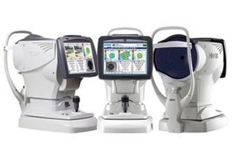
AUTO REFRACTORS
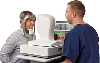
Fast, Non-Invasive Imaging
The Avanti Widefield OCT offers state-of-the-art imaging from the cornea to the choroid with exclusive technology that will change your approach to disease diagnosis and management. Quickly visualize your patient’s microvasculature with a non-invasive, dye-free technique that can be accomplished by your trained technician in a matter of seconds. With AngioVue, you can image your patients as often as need to closely follow disease progression and treatment response. Unlike traditional invasive angiography approaches, there is no need for advance procedure scheduling because OCTA information is available as you perform the OCT scan.
in OCT
The iVue®80 OCT is a powerful clinical tool that transforms the way you assess the retina, optic disc and the cornea. Quantify the thickness of the retina, nerve fiber layer, ganglion cell complex and the cornea. Track change and predict trends in RNFL and GCC thickness and precisely measure angles to aid in disease diagnosis.
Accessible and Affordable OCT Solutions
- Maximize Clinical Utility with Retina, Optic Disc and Anterior Segment Applications
- Create the System You Need with OCT + Fundus Camera Option
- Enhance Eye Health Exams and Generate New Revenue with the Exclusive iWellness Scan
in OCT
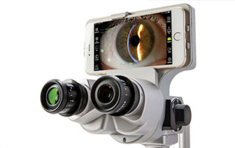
Utilizing the world’s best known and most user friendly OS. Simple but powerful proprietary slit lamp imaging app designed by ophthalmic photographers. Easily capture high resolution video or still images. Diagnose, image, integrate & educate in seconds. Update software via the App Store. Simply start the App and you’re ready to capture.
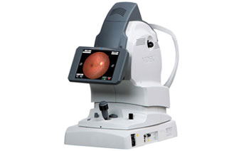
The AFC-330 delivers unsurpassed ease of use with advanced features that enhance the management of retinal disease, such as glaucoma and diabetic retinopathy. The AFC-330’s automated functions forge new ground in fundus imaging technology with focus on capturing the perfect picture every time, regardless of operator experience or skill level. The AFC-330 makes numerous command calculations per second. Only this level of automation can account for the speed of operation and accuracy of this camera – the essential foundation of practice efficiency.
Being equipped with this level of sophistication, the AFC-330 is able to align and automatically switch from anterior to posterior focusing.
in Photography
in Photography
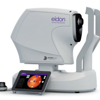
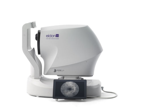
iCare EIDON is the first TrueColor Confocal system that combines the best features of Scanning Laser Ophthalmoscopy (SLO) systems with those of standard fundus imaging to set new performance standards in retinal imaging. It’s the perfect retinal imaging system that provides TrueColor and widefield views in multiple imaging modalities. iCare EIDON provides unsurpassed image quality and a unique, live, confocal view of the retina in a dilation-free operation.
Offering the best of confocal technology, iCare EIDON guarantees increased image sharpness, better optical resolution, higher details and greater contrast that takes retinal diagnostics to the next level. This combined with its easy-to-use interface and patient-friendly features make the iCare EIDON a valuable and efficient tool in any clinical setting.
in Photography
iCare DRSplus confocal fundus imaging system uses white LED illumination to offer high-quality TrueColor images. TrueColor Confocal Technology, which is considered a standard of high image quality, provides detail-rich images with greater image sharpness, optical resolution and contrast when compared to traditional fundus camera imaging.
The fast and fully automated iCare DRSplus permits imaging through pupils as small as 2.5 mm, without need of dilation, ensuring a comfortable patient experience. This easy to use device offers the advantage of quick examination time and helps speed up workflow at clinics.
in Photography
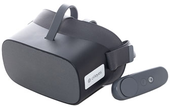
A Unique Multi-Test Platform, Not Just a Perimeter
The VisuALL VRP is a platform and not a single test device. The power of the platform is only growing as more and more capabilities are being added. It is designed to Increase Productivity and the Bottom Line
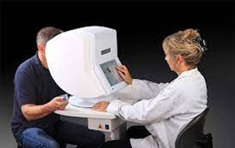
Octopus 600
The Octopus 600 covers all your essential perimetry needs ion an easy to use device: central field static testing, rapid screening, an easy to read analysis software and the ability to network it with integrated EyeSuite software.
- With its comprehensive test library for central tests including G, 32, 30-2, 24-2, M, and 10-2 and its flexible printouts both in Octopus and HFA-format it covers your essential clinical needs.
- The Octopus 600 offers the most commonly used static tests
- All Octopus perimeters offer the standard 7-in-1 printout with its well-known representations, a customizable 4-in-1 printout, a serial printout and much more.
- All Octopus perimeters allow you to import your historic HFA data.
- Use your Octopus 600 as a fast screening device and quickly distinguish between normal and abnormal visual fields. Screening can be performed with both standard white-on-white or the patient-friendly Pulsar perimetry designed for early glaucoma detection.
- Use the new Glaucoma Screening Test GST to distinguish between normal and abnormal visual fields in less than one minute.
- A screening test has to be easy to complete by definition. That’s why the patient-friendly Pulsar stimulus is recommended for screening purposes. It has been developed for early glaucoma detection and shows a short learning curve and low test-retest variability.
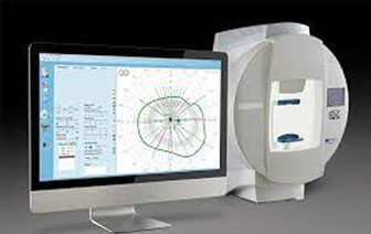
Octopus 900
Everything you can ask of a perimeter is offered in this device: full field static and kinetic perimetry, an easy to read analysis software and the ability to network it with integrated EyeSuite software.
- With its comprehensive test library for central and peripheral tests and its flexible printouts both in Octopus and HFA-format it covers all your clinical needs.
- The Octopus 900 offers all commonly used standard static tests, And if you’re still missing a test, why not create it yourself with the Custom Test option?
- Standard Octopus representations
- Smooth transition from HFA
- Get the most out of your glaucoma visual field with the highly sensitive Cluster Analysis, the intuitive Polar Analysis for structural comparisons and the easy-to-interpret EyeSuite Progression Analysis.
- Enjoy the characteristics of the manual Goldmann perimeter in a modern device with automatic electronic data filing.
The Phoenix Meibography Workstation with Topography gives a dynamic view of non-invasive tear film break-up time with tear meniscus height imaging and measurement. Other features include:
- Interference Pattern Imaging of Lipid Layer
- Imaging of the Tear Film Dynamics (Video)
- Fluorescein Imaging
- Meibomian Glands Imaging & Analysis
- Corneal Topography
- Contact lens fitting with Autofit
- Keratoconus screening with classification
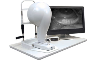
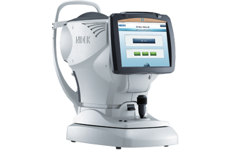
The OPD-Scan III is an autorefractor, keratometer, pupillometer, corneal topographer, and integrated wavefront aberrometer. The OPD-Scan III completes 20 diagnostic metrics in less than 10 seconds per eye. Easy alignment and automatic capture of wavefront aberrometry data ensures accurate readings. Wavefront aberrometry data is gathered from available zones up to a 9.5mm area, adding the capability to provide for calculation of mesopic refractions. Blue light, 33 ring, placido disc topography is gathered in one second. Mapping methods include OPD, visual acuity corneal topography/topographer, and more.
The OPD-Scan III Visual System is an aberrometer providing optimal and complete optical system analysis with detailed measurement reports. The easy-to-understand reports displayed on a tablet, iPhone, or laptop allow simple explanation of examination results. This unit opens a variety of business possibilities as a new communication tool.
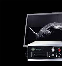
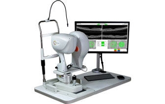
The ultimate in ultrasound performance
With customized configuration of A-Scan and B-Scan modes, Eye Cubed™ covers all your diagnostic ultrasound needs for both the posterior and anterior segments. Pre-op or post-op, A-Scan or B-Scan, retina or anterior segment: whatever your focus, Eye Cubed™ shows you more, in more detail, than any other device of its kind.
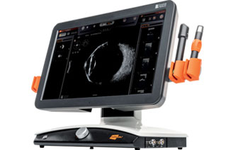
The principle is to emit alternating ultrasounds by 5 concentric transducers located in a single probe. The images thus obtained are spectacular as the entire eye is now visible with an exceptional level of detail.
- Increased depth of field allowing the visualization of the entire eye
- Outstanding resolution from the anterior part of the vitreous to the wall
- New UBM imaging technology allowing different imaging modes
- IMUv ™ exclusive motion sensor allowing to locate the ultrasound beam in real-time in the ocular diagram
- Intuitive and user-friendly software for a better navigation experience
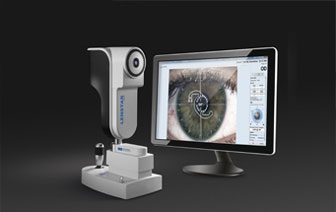
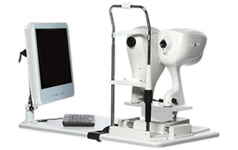
Complete optical biometry for better outcomes.
The all-in-one cataract planning platform.
Spectacle independence is the goal of cataract surgery today, Lenstar provides all the data and state-of-the-art IOL formulae required to achieve this!
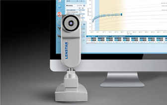
Lenstar Myopia is based on the proven Lenstar 900 technology and includes the corresponding EyeSuite software, EyeSuite Myopia. For early detection and state of the art myopia management for east educations and consultation of patients and parents.

HealthxMD is a third-party diagnostic service provider with a mission to promote a quality of vision and quality of life for the patients you see daily. They integrate transcranial doppler diagnostic testing in your office with no upfront costs.
Everything from Certified Ultrasound Sonographers to equipment and program support staff are provided by HealthxMD. You do not have to do any extra work other than serving those patients with the same quality of care delivered every day in your clinic.
in Doppler Imaging Program
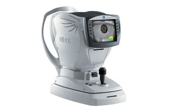
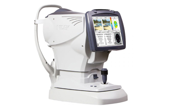
ARK-1a & ARK-1
- SLD technology
- Double ring technology
- Confidence index indicator
- Automatic pupil (2mm min.) and cornea size
- Sphere measurement range -30D to +25D
- Cyl 0.00D to ±12D
- Auto X, Y, Z eye tracking
- Accommodation
- Retro-illumination
- Tilt, LCD Screen
ARK-1s
- SLD technology
- Double ring technology
- Confidence index indicator
- Automatic pupil (2mm min.) and cornea size
- Sphere measurement range -30D to +25D
- Cyl 0.00D to ±12D
- Auto X, Y, Z eye tracking
- Glare Testing
- Low Contrast Testing
- Visual acuity chart –unaided/aided/glasses/near
- Subjective sphere refinement
- Accommodation
- Retro-illumination
- Tilt, LCD Screen
ARK-F
- SLD technology
- Double ring technology
- Automatic pupil (2mm min.) and cornea size
- Sphere measurement range -30D to +25D
- Cyl 0.00D to ±12D
- Auto X, Y, Z eye tracking
- Accommodation
- Retro-illumination
- Tilt and Swivel LCD Screen
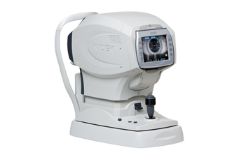
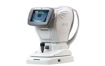
Tonoref II ARK with Tonometry
- Automatic Refraction
- Automatic Keratometry
- Non-Contact Tonometry
- Space Saving
- Time Saving
- High-Speed Measurements
- Accurate Data
- EyeTracking System
- Auto Measure
- Adjustable Monitor
- Motorized Chinrest
- Softer Air Puff
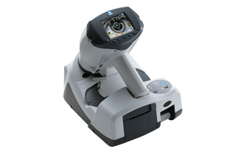
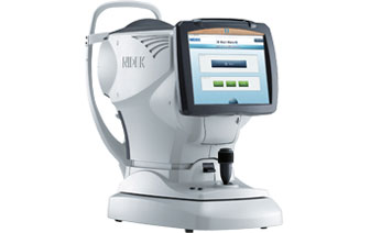
HandyRef-K Portable ARK
- Compact Form Factor
- Pupil Zone Imaging Method
- Super Luminescent Diode (SLD) & Highly Sensitive CCD
- SynchroScan Technology
- Full Graphic LCD with 3.5-inch Color Screen
- Achieve Precise Measurements Anytime, Anywhere.
- Cataract Measurement Mode
- Quick Measurement Mode
- Auto Measure
- Keratometry Measurement with Mire Ring
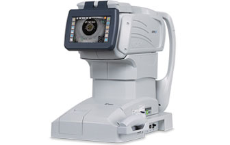
OPD III
- Wavefront aberrometry with 2,520 light vector data points
- Corneal topographer/topography with 11,880 data point mapping
- Retro-illumination
- Day/Night Rx
- Auto X, Y, Z eye tracking
- Discerns AR vs. WF patients and their starting refraction point
- Sphere measurement -20D to +22D. Cyl 0.00D to ±12D
- Large, tilt LCD screen for superior viewing angle and operational position options
OPD III Visual System
- Wavefront aberrometry with 2,520 light vector data points
- Corneal topographer/topography with 11,880 data point mapping
- Retro-illumination
- Day/Night Rx
- Auto X, Y, Z eye tracking
- Discerns AR vs. WF patients and their starting refraction point
- Sphere measurement -20D to +22D. Cyl 0.00D to ±12D
- Large, tilt LCD screen for superior viewing angle and operational position options
Ready to Learn More?
If you want to learn more about any of the instruments you see here, we are happy to speak with you! Click below to get in touch with Scott or Drew or give either of us a call!