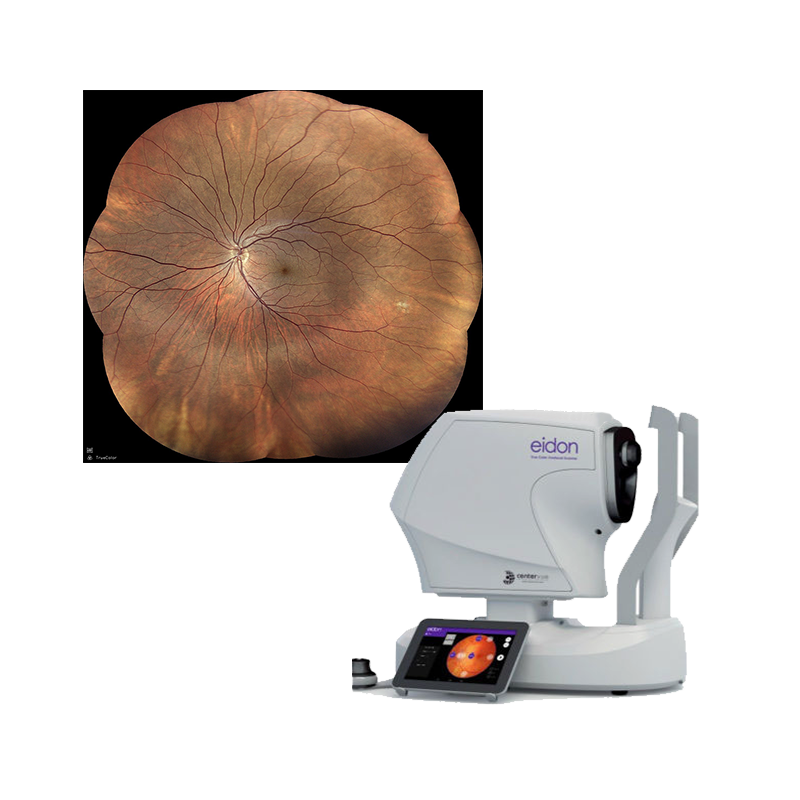
Eidon
iCare EIDON widefield TrueColor confocal fundus imaging system
iCare EIDON is the first TrueColor Confocal system that combines the best features of Scanning Laser Ophthalmoscopy (SLO) systems with those of standard fundus imaging to set new performance standards in retinal imaging. It’s the perfect retinal imaging system that provides TrueColor and widefield views in multiple imaging modalities. iCare EIDON provides unsurpassed image quality and a unique, live, confocal view of the retina in a dilation-free operation.
Offering the best of confocal technology, iCare EIDON guarantees increased image sharpness, better optical resolution, higher details and greater contrast that takes retinal diagnostics to the next level. This combined with its easy-to-use interface and patient-friendly features make the iCare EIDON a valuable and efficient tool in any clinical setting.
Key features
- Multiple imaging modalities including TrueColor, blue, red and Red-Free and infrared confocal images
- Widefield, ultra-high-resolution imaging up to 200°
- Capability to image through cataract and media opacities
- Dilation-free operation (minimum pupil 2.5 mm)
- Flexibility of fully automated and fully manual mode
- All-in-one compact design, no additional PC required

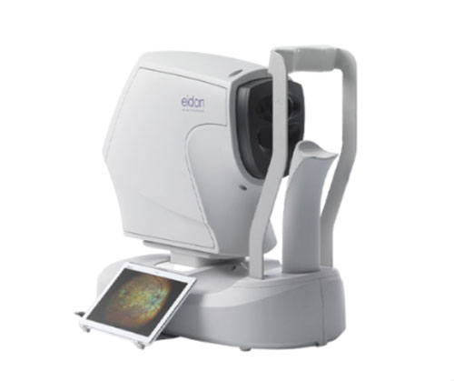
Eidon AF
iCare EIDON AF blue autofluorescence confocal fundus imaging system
iCare EIDON AF TrueColor confocal scanner offers the best of iCare EIDON technology with the added advantage of autofluorescence imaging capabilities. The fully automated scanner captures high resolution, accurate imaging using multiple modalities — TrueColor, red and blue imaging, Red-free , infrared and blue autofluorescence. The cutting-edge device obtains excellent images in pupils as small as 2.5 mm. The quick and non-invasive autofluorescence imaging technique improves patient comfort and experience. With a fully computer-assisted acquisition mode, any staff member can run tests, which can help reduce waiting time and aid in improving workflow efficiency. This device truly offers the best to both patients and eyecare professionals.
Key features
- TrueColor, blue autofluorescence, infrared and RGB channels confocal images
- Panoramic view of the retinal autofluorescence (up to 110°)
- High details and contrast in the same device
- Short exam time and enhanced patient comfort
- Easy to use, speeds up the patient workflow
"I was very impressed with the high level of image quality... great resolution. I feel like I’m seeing smaller retinal blood vessels than I could ever imagine with my previous camera."

Dr. Jesse Jones
Owner & Lead Optometrist, East Falls Eye Associates

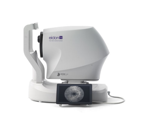
Eidon FA
iCare EIDON FA fluorescein angiography confocal fundus imaging system
iCare EIDON FA is a top-end imaging system that combines the best of the iCare EIDON technology with automatic Fluorescein Angiography capability to provide a complete suite of imaging modality of unsurpassed image quality. It offers widefield imaging of Fundus Fluorescein Angiography while preserving the image quality, sharpness, and details, even in the periphery.
iCare EIDON FA also offers the added advantage of capturing a detailed ultra-high resolution FA video recording which provides a realistic and dynamic view of retinal vasculature and circulation mechanisms that may be missed with static flash photography
Key features
- Includes all the features and functionalities of EIDON technology
- Complete suite of fundus imaging capabilities
- High resolution Fluorescein Angiography images
- High resolution and dynamic Fluorescein Angiography video view
- TrueColor confocal technology for high-quality, accurate imaging
- Fully automatic, easy to use and requires minimal staff training

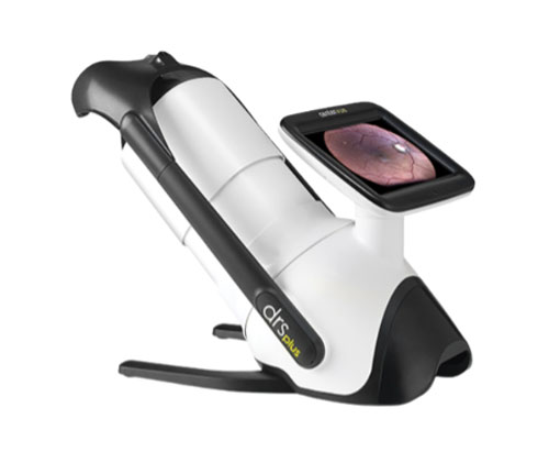
DRS PLUS
iCare DRSplus TrueColor confocal fundus imaging system
iCare DRSplus confocal fundus imaging system uses white LED illumination to offer high-quality TrueColor images. TrueColor Confocal Technology, which is considered a standard of high image quality, provides detail-rich images with greater image sharpness, optical resolution and contrast when compared to traditional fundus camera imaging.
The fast and fully automated iCare DRSplus permits imaging through pupils as small as 2.5 mm, without need of dilation, ensuring a comfortable patient experience. This easy to use device offers the advantage of quick examination time and helps speed up workflow at clinics.
Key features
- TrueColor Confocal Technology
- Multiple imaging modalities including Red-free, external eye and stereo view imaging
- 2.5 mm minimum pupil size
- Fast, easy and fully automated operations
- Mosaic function creates retinal panoramic views up to 80°
- Remote Viewer allows for reviewing from devices on the same local area network
- Remote Exam feature enables executing an exam from a distance

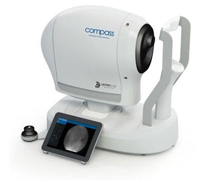
Compass
iCare COMPASS automated perimeter with active retinal tracking
iCare COMPASS combines visual field tests, fixation loss correction by a real-time retinal tracker and confocal TrueColor fundus imaging taking visual field analysis to the next level. With touch screen, auto-alignment, non-mydriatic, easy-to-disinfect, and trial lens-free operation, iCare COMPASS is patient-friendly and easy to use, saving time and helping in improving clinical performance.
iCare COMPASS is the first automated perimeter that can perform standard visual field tests using a real-time retinal tracker while delivering ultra-high resolution confocal TrueColor fundus images at the same time.
Key features
- Standard automated perimetry
- Active retinal tracking compensating for poor patient fixation in real-time
- Auto-focus — no trial lens needed
- Hygienic design
- Illustrative fixation analysis; fixation area and plot
- High-resolution confocal TrueColor imaging of the retina
- No dilation of pupil needed, the patient can blink freely and the test can be suspended at any time without data loss
- Ease of use & minimal operator training


